Anatomy Rib Cage Posterior View - Human Skeletal System Rib Cage Anatomy Ilustración de ... / The upper 7 ribs on each side of the cage connect distally.
Anatomy Rib Cage Posterior View - Human Skeletal System Rib Cage Anatomy Ilustración de ... / The upper 7 ribs on each side of the cage connect distally.. The rib cage is formed by the vertebral column, ribs, and sternum and encompasses the heart and lungs. The rib cage is an arrangement of bones in the thorax and vertebrates. Anterior and posterior view of. 5.5 ribs right ribs, superior view. The costotransverse ligaments in human:
Lateral flexion of the rib cage at the vertebral joints (continued). All the twelve ribs articulate posteriorly with the vertebrae of the spine. This muscle is present posteriorly within the thoracic wall. Bones and joints of the thorax. Abdominal viscera anatomical location posterior view.
3D Skeletal System: 7 Interesting Facts about the Thoracic ... from info.visiblebody.com The rib cage is formed by the vertebral column, ribs, and sternum and encompasses the heart and lungs. Rib bones are similar, both have spongy bone). This muscle is present posteriorly within the thoracic wall. Abdominal viscera anatomical location posterior view. The vertebral column is in neutral position. Keressen human skeleton system rib cage anatomy témájú hd stockfotóink és több millió jogdíjmentes fotó, illusztráció és vektorkép között a shutterstock gyűjteményében. It is split into superior and inferior fibres. Human skeleton ribs vertebral column anatomy royalty free.
In humans, the rib cage, also known as the thoracic cage, is a bony and cartilaginous structure which surrounds the thoracic cavity and supports the pectoral girdle (shoulder girdle), forming a core portion of the human skeleton.
Anatomical illustrations of the thoracic cage and the mammary gland. Abdominal viscera anatomical location posterior view. For more anatomy content please follow us and visit our website: 5.5 ribs right ribs, superior view. Bones and joints of the thorax. The rib cage is an arrangement of bones in the thorax and vertebrates. Human skeleton system rib cage posterior view anatomy. The rib cage is formed by the vertebral column, ribs, and sternum and encompasses the heart and lungs. Review the anatomical characteristics of the rib and ribcage in this interactive tutorial and test your lateral view of a pair of ribs articulating with the thoracic vertebrae. Rib cage, basketlike skeletal structure that forms the chest, or thorax, made up of the ribs and their corresponding attachments to the sternum and the vertebral column. Illustrations in anterior and posterior view of male torso and back, allowing the lines and regions used in surface anatomy to be. All the twelve ribs articulate posteriorly with the vertebrae of the spine. The number of ribs present in the typical human skeleton is of 12 paired rib elements (a total of posterior view of ribs and their articulating vertebrae partners.
Rib cage illustration stock photos rib cage illustration. The upper 7 ribs on each side of the cage connect distally. Anatomical illustrations of the thoracic cage and the mammary gland. Crossfit shoulder muscles part 2 posterior musculature. The ribs are curved, flat bones which form the majority of the thoracic cage.
Posterior Rib Anatomy - Anatomy Diagram Book from accessmedicine.mhmedical.com Each rib forms two joints the ribs are a set of twelve paired bones which form the protective 'cage' of the thorax. Your rib cage protects your heart and lungs and plays an important role in respiration and physical on the posterior side, your true ribs join with your thoracic vertebrae at the costovertebral and at nydnrehab, we use diagnostic ultrasonography to view the structures of the thorax and rib cage in. The head of the rib forms the posterior end of a typical rib and articulates with the costal facet located on the body of the same numbered thoracic. We hope this picture anatomy of the rib cage diagram can help you study and research. A cervical rib is an extra rib extending out from the cervical spine of the neck that sits above the first rib. The rib cage is an arrangement of bones in the thorax and vertebrates. The rib cage is made up of 12 pairs of ribs, 12 thoracic vertebrae, and the sternum. Human skeleton system rib cage anatomy posterior view.
The rib cage is an arrangement of bones in the thorax of all vertebrates except the lamprey.
Human skeleton system rib cage anatomy posterior view. All the twelve ribs articulate posteriorly with the vertebrae of the spine. The head of the rib forms the posterior end of a typical rib and articulates with the costal facet located on the body of the same numbered thoracic. the rib cage has 12 sets of ribs. The ribs are curved, flat bones which form the majority of the thoracic cage. 5.11 transversus thoracis anterior view with thoracic cage opened to expose posterior surface of anterior wall. Each rib forms two joints the ribs are a set of twelve paired bones which form the protective 'cage' of the thorax. Human skeleton ribs vertebral column anatomy royalty free. Crossfit shoulder muscles part 2 posterior musculature. For more anatomy content please follow us and visit our website: It is important to note that both the posterior and anterior articulations. The part of the muscle is thought to depress the ribs. Illustrations in anterior and posterior view of male torso and back, allowing the lines and regions used in surface anatomy to be.
It is split into superior and inferior fibres. Anterior and posterior view of. the rib cage has 12 sets of ribs. Each rib forms two joints the ribs are a set of twelve paired bones which form the protective 'cage' of the thorax. Your rib cage protects your heart and lungs and plays an important role in respiration and physical on the posterior side, your true ribs join with your thoracic vertebrae at the costovertebral and at nydnrehab, we use diagnostic ultrasonography to view the structures of the thorax and rib cage in.
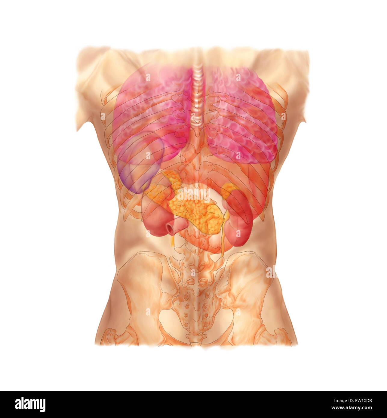
Abdominal quadrants, posterior view with internal organs ... from c8.alamy.com It is split into superior and inferior fibres. The rib cage surrounds the lungs and the heart, serving as an important means of bony protection for these vital organs. (movement can also be of the figure leaning toward the left.) For more anatomy content please follow us and visit our website: The rib cage is the arrangement of ribs attached to the vertebral column and sternum in the thorax of most vertebrates, that encloses and protects the vital organs such as the heart, lungs and great vessels. The rib cage is an arrangement of bones in the thorax and vertebrates. This muscle is present posteriorly within the thoracic wall. The shaded areas indicate the extent of the pleural cavities not filled by the lungs.
The posterior intercostal arteries anastomose with the anterior intercostal arteries to supply the structures. The outer border is convex, thick, and rounded, and at its posterior part gives attachment to the first. The illustrations were drawn in adobe illustrator using data from medical imaging surface anatomy: Structure of a typical rib: It is split into superior and inferior fibres. The shaded areas indicate the extent of the pleural cavities not filled by the lungs. The rib cage is an arrangement of bones in the thorax and vertebrates. The costotransverse ligaments in human: They articulate with the vertebral column posteriorly, and terminate anteriorly as cartilage (known as costal. Cureus an unusual back muscle identified bilaterally case. Human skeleton system rib cage posterior view anatomy. Bones and joints of the thorax. Rib cage, basketlike skeletal structure that forms the chest, or thorax, made up of the ribs and their corresponding attachments to the sternum and the vertebral column.
The rib cage is an arrangement of bones in the thorax of all vertebrates except the lamprey anatomy rib cage. Projection on the rib cage of the heart, lungs and diaphragm.


Posting Komentar
0 Komentar